Spinal compression fracture · Diabetes
Your bone density scan came back normal. Diabetic bone still suffers a compression fracture, and the scan cannot see why.
A vertebral compression fracture in someone with type 2 diabetes is often a fracture the DXA scan said could not happen. We treat the pain. We fix the fracture when it needs fixing. And we go after the bone chemistry the scan cannot measure.
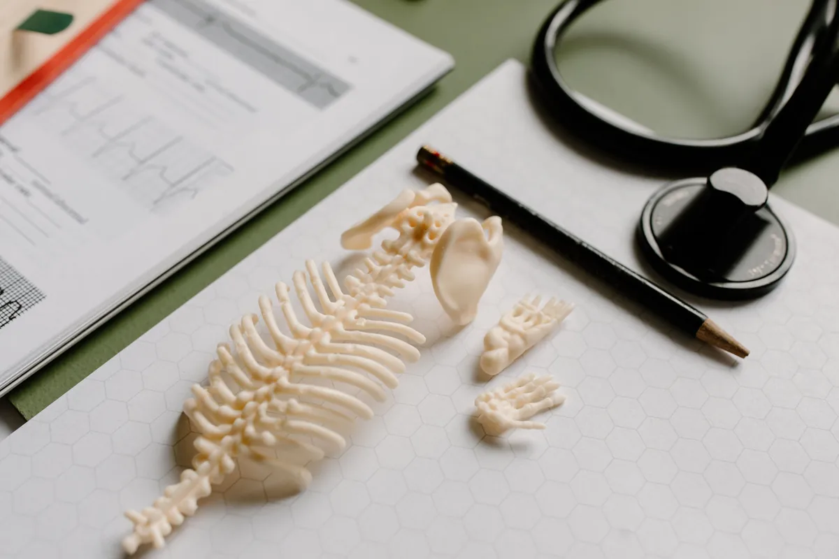
Why can you get a compression fracture with a normal bone density scan?
A DXA scan counts mineral. It reports how much calcium is packed into a square centimeter of bone. For most osteoporosis, that number tracks fracture risk well. Type 2 diabetes breaks the link. A meta-analysis of the published cohorts found that people with type 2 diabetes have higher bone density than people without it. They still break their hips more often. In type 1 the risk is several times higher. The mineral is there. The bone fails anyway, because mineral is not what makes bone tough.
Toughness comes from collagen, and high glucose ruins collagen. Sugar bonds to the protein fibers and cross-links them, the way old rubber hardens. The bone turns stiff and brittle instead of springy. At the same time, long-term high insulin and inflammatory signals hold back the cells that build new bone. Daily life makes tiny cracks in bone, and now they get patched more slowly. Marrow fat spreads into space that should hold bone-building cells. None of that shows on a density scan. That is why a person can be told their bones are fine in March and break a vertebra reaching for a hose in July.
Then the third driver. Thirty years in a desk chair or a cab seat removes the loading that tells bone to rebuild. An indoor shift removes the sunlight that makes vitamin D. Steroid injections for an unrelated knee or shoulder add up to a glucocorticoid dose the bone has to absorb. The fracture is a metabolic and a behavioral event before it is a mechanical one. Treat it as a cracked bone alone, and the next fracture is already scheduled.
What does a compression fracture feel like?
Sudden pain in the middle of the back, at one spot you could cover with your palm. It comes after a load that should not have mattered, or after nothing at all. It is worse standing and walking and better lying flat. It can wrap around the ribs in a band. Many are called a muscle strain at the first visit because no X-ray was taken. The front of the vertebra collapses, the spine tips forward, and the load on the next vertebra rises. That is why a first fracture predicts a second. It is also why the pain sometimes outlasts the healing. The joints behind the wedge now carry weight they were never built for. That is facet pain with a different origin story.
NOT A CLINIC APPOINTMENT
Weakness or numbness moving down the legs. Any change in bladder or bowel control. Fever or drenching night sweats with the back pain. A history of cancer, or weight you did not try to lose. A fracture in someone under fifty with no reason to have thin bone. Pain that is the same lying down as standing up. These point to something other than a simple osteoporotic collapse. The emergency page says where to go.
Do kyphoplasty and vertebroplasty work for compression fractures?
Two 2009 trials in the New England Journal of Medicine compared vertebroplasty with a sham procedure and found no difference. They took fractures up to a year old, many with moderate pain. The sham included local anesthetic on the bone surface, which is itself a treatment. Two later trials sharpened the question. VAPOUR, in 2016, took only fractures under six weeks old with pain of at least 7 out of 10. Most were in hospital patients. Vertebroplasty beat sham at two weeks and at six months. VERTOS IV, in 2018, took fractures up to nine weeks old and used the same anesthetic-on-bone sham. Both groups got much better, with no difference between them at a year. The FREE trial compared balloon kyphoplasty with non-surgical care in 300 patients with fractures under three months old. It found faster improvement in the kyphoplasty group at one month, and the gap narrowed by twelve.
Read together, the trials say something specific. Cement helps when the fracture is fresh and the pain is severe and in one spot. The MRI must show the bone is still edematous (still swollen with fluid). And the other choice is weeks in bed. It does not help a fracture that healed last year. It does not help pain that has moved to the joints behind it. That is our position. It is why the MRI comes before the decision, not after it.
KYPHOPLASTY, WHEN IT FITS
A balloon restores some of the lost height and makes a cavity. Cement fills it and steadies the fracture the same day. The kyphoplasty page covers the procedure and its recovery rules. Two things to know now: this is one of the few procedures here done under mild sedation, so you will need a driver; and for an Illinois patient it can be scheduled in a hospital setting, including at Anderson Hospital in Maryville, where Dr. Gurpreet Singh Padda, MD, MBA, MHP holds full privileges.
How is a compression fracture treated, and how do you prevent the next one?
With or without cement, the fracture gets a short window of bracing and pain relief, and a plan for ending it. Opioids, if they are needed at all, are a bridge measured in days. The taper is written on the first day; this page explains why. The bloodwork is the part most people did not expect. We check vitamin D, calcium and parathyroid hormone, thyroid, and testosterone where it matters. We check the A1C and fasting insulin that tell us whether the collagen problem above is active. If the numbers call for a bone-building drug, endocrinology writes that prescription and we work with them.
The rest of the plan decides whether there is a next fracture. Bone rebuilds in response to strain, sensed by cells inside it. So we prescribe resistance loading, not walking alone. We prescribe protein, because sarcopenic wasting (muscle loss) and fragile bone travel together. We prescribe sleep, because bone turnover runs on a nightly clock. And Acceptance and Commitment Therapy is delivered in-house by a licensed, pain-trained behavioral clinician. Here is why. The most common outcome after a vertebral fracture is fear of movement. Then comes deconditioning. Then comes the fall that causes the second fracture. That work is 40 to 50 percent of the protocol here.
Getting here with a back that will not sit
An acute fracture makes the car the hardest part of the appointment, so we plan the order. Imaging comes first if you do not already have it. Then one consultation decides the plan. Then comes the procedure day, if there is one. From Alton it is about 30 minutes without traffic by way of the Clark Bridge and I-270. From Edwardsville, it is about 40 by I-270 or I-55 and I-70. Tell us when you call that sitting is the problem. We schedule around it.
Frequently asked questions
Do I need a referral for a compression fracture?
No. You can call and book the visit yourself, and a fresh fracture is seen quickly. Some insurance plans want a referral on file before they pay, so check your card. Diabetic bone breaks more easily than a scan suggests, which why a pain clinic checks your blood sugar explains.
How long after a compression fracture can kyphoplasty still help?
The trials that showed benefit took fractures measured in weeks, not months. The MRI is the judge. Bone that still shows edema (fluid) is still a candidate. Bone that has healed solid is not. The kyphoplasty page walks through how that decision is made.
Does the cement prevent the next fracture?
It steadies the one it is in. The next fracture is decided by collagen quality, bone turnover, muscle and balance. That is why the metabolic work and the loading program are the real prevention.
Why does my back still hurt months after the fracture healed?
A wedged vertebra changes the angle of the spine above and below it. The small joints at those levels start carrying load they were not built for. That pain answers to a diagnostic block and, when confirmed, to radiofrequency ablation, not to more cement.
Will I need someone to drive me?
For kyphoplasty, which uses mild sedation, a driver is required. For the consultation, the diagnostic blocks and most other procedures here there is no sedation and most people drive themselves; what to expect lays out the day.
How serious is a compression fracture of the spine?
More serious than the pain alone suggests. The front of the vertebra collapses, the spine tips forward, and the load on the next vertebra rises. That is why a first fracture predicts a second. The pain can also outlast the healing, because the joints behind the wedge now carry weight they were not built for. Weakness or numbness down the legs, or any change in bladder or bowel control, is an emergency, not a clinic visit.
How is a compression fracture diagnosed?
With imaging, not a guess. Many are called a muscle strain at the first visit because no X-ray was taken. We get imaging first if you do not already have it. The MRI shows whether the bone is still swollen, which decides whether cement can help. Bloodwork follows: vitamin D, calcium, parathyroid hormone, thyroid, A1C and fasting insulin. In type 2 diabetes the density scan can read normal while the bone is fragile.
What should you not do after a compression fracture?
Do not treat it as a cracked bone and nothing more, or the next fracture is already scheduled. Do not let fear of moving turn into weeks of sitting still. Deconditioning sets up the fall that causes a second fracture. Do not count on walking alone to rebuild bone, because bone responds to resistance loading. And opioids, if they are needed at all, are a bridge measured in days, with the taper written on the first day.
Is sitting bad for a compression fracture?
Upright positions load the broken vertebra. So the pain is usually worse standing and walking and better lying flat. The car ride is often the hardest part of an appointment. Tell us when you call that sitting is the problem. We plan the visits around it: imaging first if you need it, then one consultation that decides the plan, then the procedure day if there is one.
Related reading
Sources
- Vestergaard P. Discrepancies in bone mineral density and fracture risk in patients with type 1 and type 2 diabetes: a meta-analysis. Osteoporosis International, 2007. PubMed 17068657
- Napoli N et al. Mechanisms of diabetes mellitus-induced bone fragility. Nature Reviews Endocrinology, 2017. PubMed 27658727
- Buchbinder R et al. A randomized trial of vertebroplasty for painful osteoporotic vertebral fractures. New England Journal of Medicine, 2009. PubMed 19657121
- Clark W et al. Safety and efficacy of vertebroplasty for acute painful osteoporotic fractures (VAPOUR): a multicentre, randomised, double-blind, placebo-controlled trial. Lancet, 2016. PubMed 27544377
- Firanescu CE et al. Vertebroplasty versus sham procedure for painful acute osteoporotic vertebral compression fractures (VERTOS IV): randomised sham controlled clinical trial. BMJ, 2018. PubMed 29743284
- Wardlaw D et al. Efficacy and safety of balloon kyphoplasty compared with non-surgical care for vertebral compression fracture (FREE): a randomised controlled trial. Lancet, 2009. PubMed 19246088
We will tell you whether cement is the right answer for your fracture, and what we do if it is not
The MRI and the bloodwork decide it, not the calendar. If your fracture is past the window, you will hear that, and you will hear what is actually generating the pain instead.
505 Buckeye Dr, Suite 200
Troy, IL 62294
Procedure clinic: 4499 Woodson Rd, St. Louis, MO 63134, off I-70 next to St. Louis Lambert International Airport. One bridge from the Metro East.